Product · By use case
Fat, fibrosis and liver zones, measured on every slide.
Fat droplet area with large and small droplets apart, collagen area on picrosirius red, fat cell size, and the zones around each vein.
A real run · H&E, mouse liver
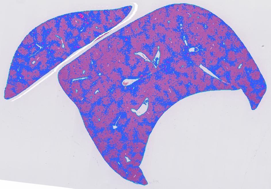
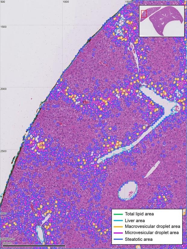
Fat in a mouse liver, measured droplet by droplet.
The request
“Steatotic area and total lipid area, as a percent of the liver. Leave out the large vessels.”
What came back, in 2 minutes
- 2.55%
- of the liver is lipid, in 25,043 droplets.
- 1.98%
- is small droplets, and 0.57% large ones.
- 13.3%
- of the liver is steatotic, estimated from the droplets.
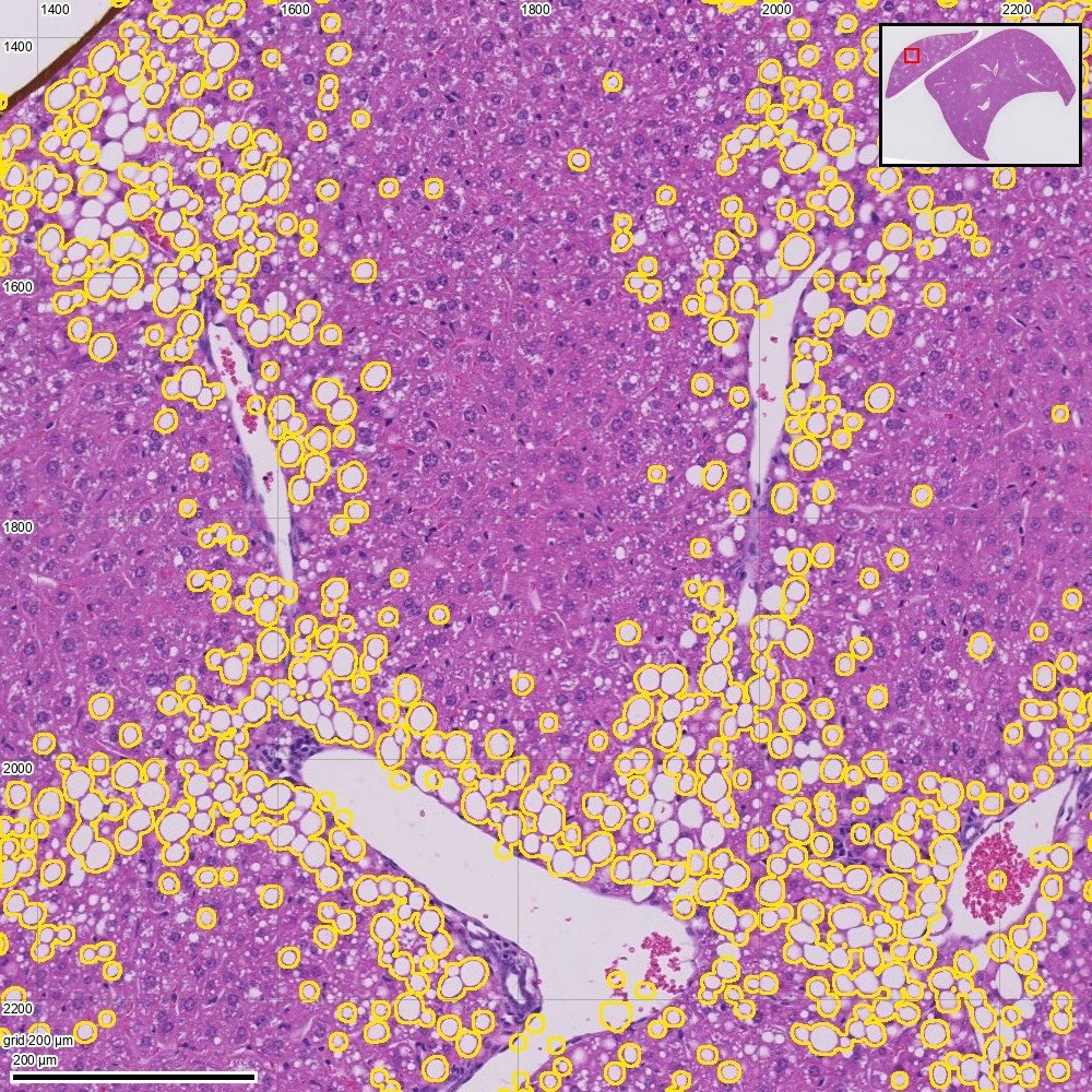
Fat droplets
Every droplet in the section outlined, then split into large and small by size.
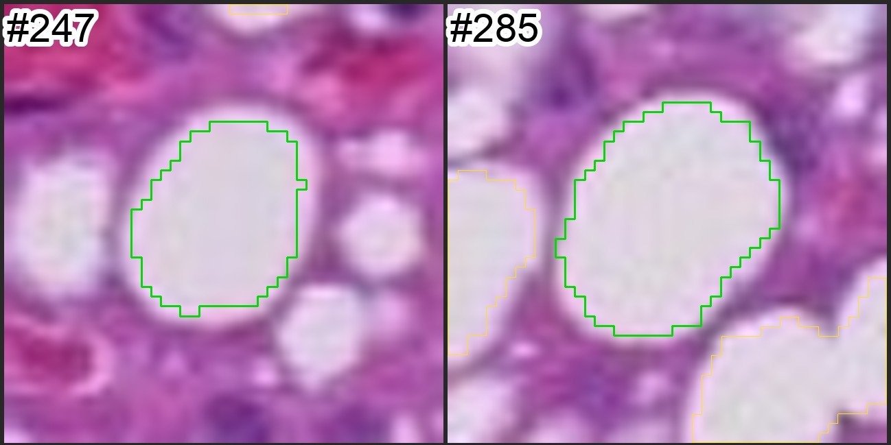
Checked up close
The AI opens droplets one by one to check each outline before it reports.
What it flagged for a person to check
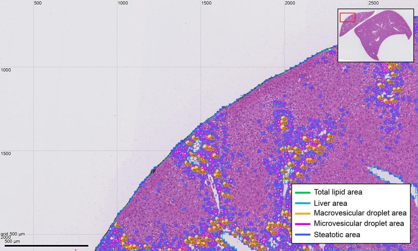
Glycogen is not fat
Lacy clear cells were kept out of the lipid. The report says this split was not checked cell by cell.
Lipid is probably undercounted
Merged droplet clusters can be missed, so the true lipid area is likely higher. The report says so.
Mouse liver, high-fat diet, H&E. Slide: Budelmann et al., 2021, CC BY 4.0.
A real run · H&E, rat liver
Zones and injury in rat liver, on plain H&E.
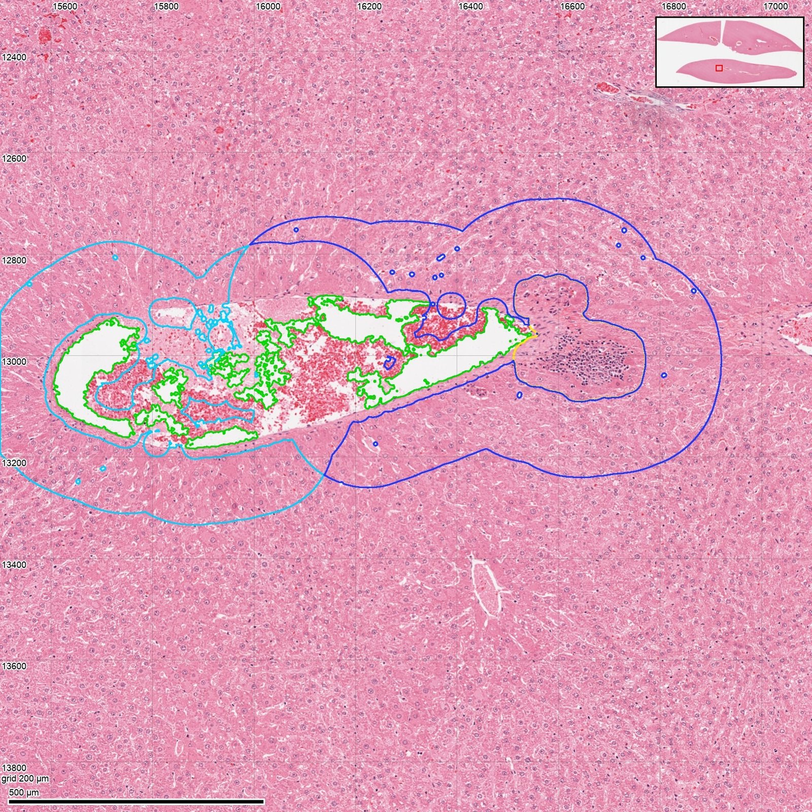
Liver zones
Rat liver after 28 days of phenobarbital. Fewer nuclei per mm² near central veins (906 against 1,352), where cells are enlarged.
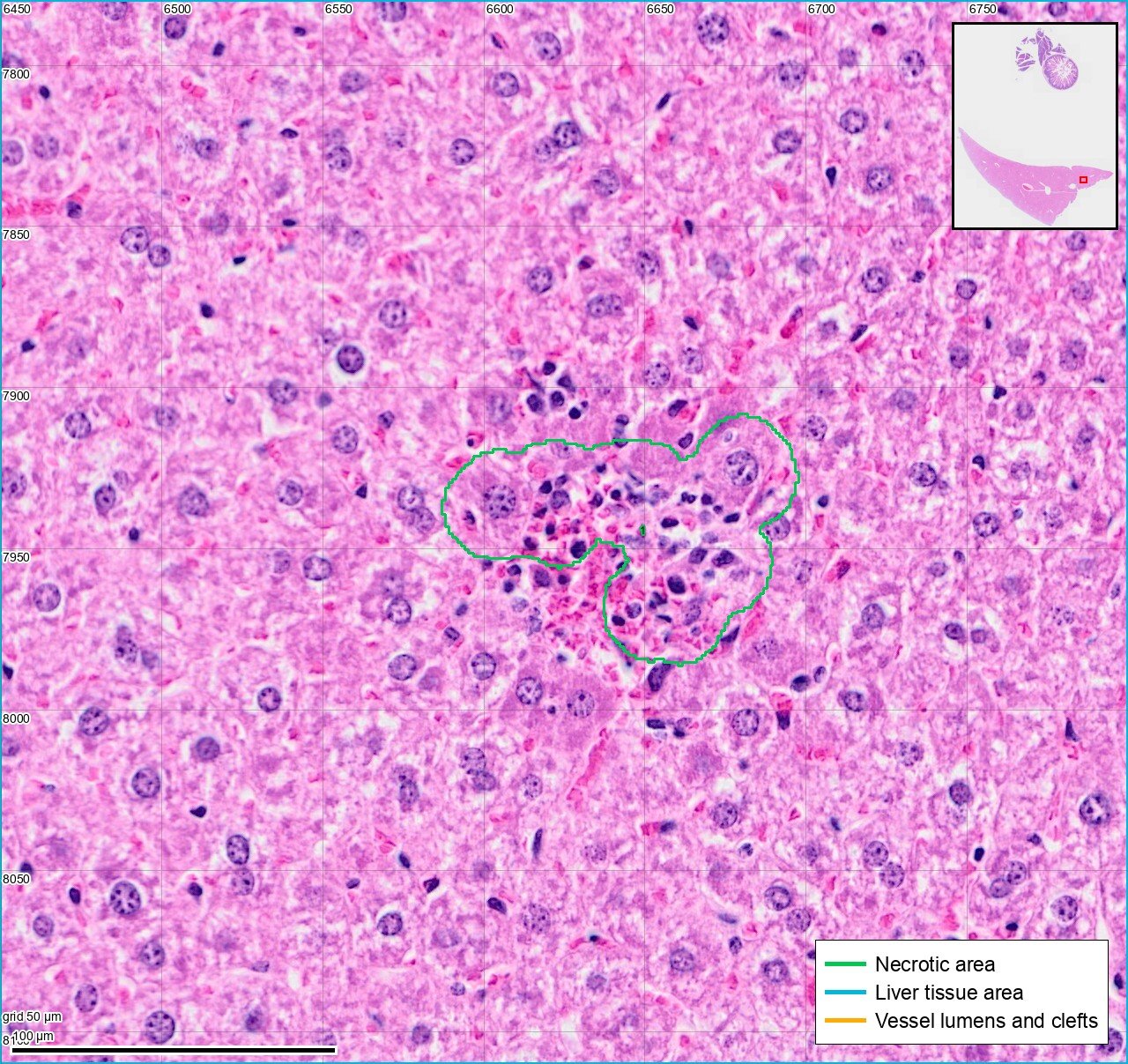
Injury
Foci of necrosis and inflammation found, outlined and measured as a percent of the liver.
What it flagged for a person to check
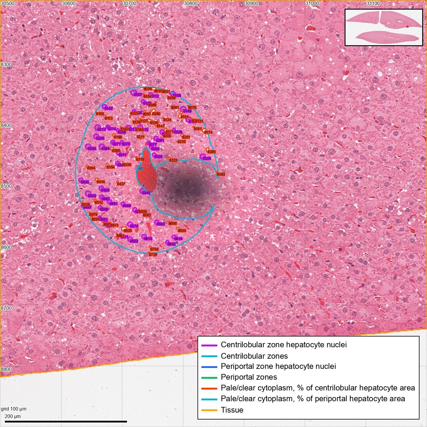
A smudge that is not nuclei
Out-of-focus grey blobs stain dark like nuclei. They were found and cut out of the liver zones.
Slides: Open TG-GATEs; Hautier et al., 2024.
What you can ask for
Ask for these, or anything else you can see.
In plain words. If it can be seen on the sample, the AI can build a method to measure it.
- Steatotic area and total lipid area
- Large and small fat droplets apart
- Collagen area on picrosirius red
- Fat cell size in adipose tissue
- Nuclei per mm² in each liver zone
How it works
You upload, and we run the study.
01
Upload your slides or flow files
Any stain or panel. One sample is enough to start.
02
Say what to measure
In plain words, the way you would ask a colleague.
03
We run the study
The AI builds the method on one sample. A person checks it and locks it, and code measures every sample with it.
04
Get the report
Numbers, outlines, gates and flags for every sample. You pay for each slide or flow file.
Why VSQRD
- Every reported number measured by code, never by eye
- First-hand research and wet-lab perspective
- Direct founder involvement from discovery to rollout
Start with one study you have already reported.
Upload its samples and tell us what you measured. We run it and show you where our numbers and yours differ.
Book a Discovery Session